URGENT UPDATE: A groundbreaking study published on July 5, 2025, reveals that a new deep learning model has transformed the 3D imaging of fruit tissues, achieving extraordinary accuracy in analyzing apple and pear microstructures. This innovative approach, developed by a team at KU Leuven led by Pieter Verboven, outperforms traditional 2D methods and offers a non-destructive, automated solution for quantifying plant tissue morphology.
The traditional microscopy techniques have long been limited by their extensive sample preparation and small field of view. Recent advancements in X-ray micro-CT imaging have opened new doors, yet challenges remain in accurately separating complex tissue features. This new deep learning framework addresses these issues head-on, providing a solution that is both efficient and precise.
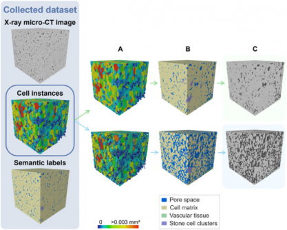
The study, featured in Plant Phenomics, showcases a 3D panoptic segmentation framework built on a novel extension of the Cellpose model and a 3D Residual U-Net. This model effectively labels and quantifies fruit tissue microstructure from native X-ray micro-CT images. Remarkably, it achieved an Aggregated Jaccard Index (AJI) of 0.889 for apples and 0.773 for pears, significantly surpassing the performance of existing 2D models.
The implications of this research are profound. For plant scientists, this model provides a powerful tool that enhances understanding of how microscopic structures influence vital processes such as water, gas, and nutrient transport. The ability to analyze cellular arrangements can lead to breakthroughs in fruit texture, storability, and even susceptibility to physiological disorders like browning.
The 3D model employs advanced techniques, including instance segmentation to differentiate individual parenchyma cells and semantic segmentation for classifying various tissue types. Evaluation metrics revealed exceptional performance, with significant metrics indicating near-perfect segmentation of pore spaces and cell matrices.
Visual validation from the study confirmed accurate detection of vascular bundles in notable apple varieties such as ‘Kizuri’ and ‘Braeburn’, along with smooth segmentation of stone cell clusters in ‘Celina’ and ‘Fred’ pears, achieving a Dice Similarity Coefficient (DSC) as high as 0.90. However, researchers noted that while additional data augmentation strategies were tested, they did not further enhance model performance, likely due to dataset imbalances.
This pioneering research, funded by the Research Foundation – Flanders, not only sets a new standard for plant anatomy studies but also creates scalable frameworks that can be applied across various crops. It opens up exciting opportunities for further exploration into tissue development, ripening, and stress responses.
As the scientific community embraces this technological advancement, the potential to integrate artificial intelligence into agricultural practices becomes clearer, promising improved efficiency and accuracy in food science research. The swift adoption of this deep learning model could fundamentally alter how researchers study and understand plant physiology, making it a pivotal development in the field.
Stay tuned for more updates as this revolutionary model gains traction in the scientific community, paving the way for faster, more precise agricultural research.